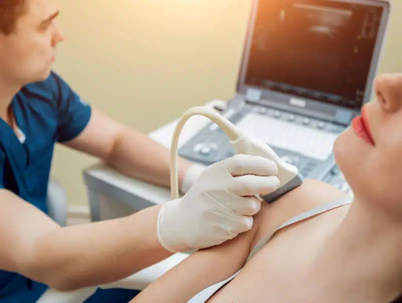
Private Breast Ultrasound Scan
Private Breast Ultrasound (Two Breast)
£259
We provide a comprehensive bilateral breast scan including axilla (underarms), assessing breast tissue and nearby lymph nodes for early signs of lumps, cysts or inflammation. This type of ultrasound scan is often used to diagnose and evaluate breast abnormalities, such as lumps, cysts, or suspicious masses found during a physical examination or a mammogram.
A breast ultrasound scan can be used to:
- Identify and evaluate breast lumps or cysts
- Determine if a breast lump is solid or filled with fluid
- Detect abnormalities in the breast tissue, such as masses or areas of increased density
- Evaluate changes in the breast tissue after surgery or other medical treatments
- Assist in guiding a biopsy or other medical procedures
Book Your Appointment
Please select a location and time slot to proceed with the booking
If you can’t make a payment online, please call our office to book your appointment. We’re here to assist you!
Tel: 020 7101 3377
Our Latest Google Reviews
EXCELLENTTrustindex verifies that the original source of the review is Google. FantasticPosted onTrustindex verifies that the original source of the review is Google. Highly recommended. A very professional and caring team. I was seen promptly over the bank holiday weekend for severe pain, and received excellent service from Dr. Salehi Very happy 😊Posted onTrustindex verifies that the original source of the review is Google. Got an appointment really quickly, service was good and I received the written email report before I got home .Posted onTrustindex verifies that the original source of the review is Google. I attended this clinic in St albans 04/04/2026. Was for a 1 limb scan of my leg. Was all very friendly and very thorough, explained everything as we went along. Would definitely recommend excellent service and got all my scans etc same day. Well worth having this done, if any concerns.Posted onTrustindex verifies that the original source of the review is Google. Absolutely amazing staff,very kind and professional!Posted onTrustindex verifies that the original source of the review is Google. I made an appointment for a scan this morning. Online booking system was very easy, giving many suggested appointment opportunities The staff were very efficient and kind. The scan was explained to me very fully by Dr Salehi whilst it took place, and the report was emailed to me within two hours Highly recommend and very good value, especially for reassurance as wellPosted onTrustindex verifies that the original source of the review is Google. I attended London Private Ultrasound on the 3rd April 2026, where Dr Fakilian, cardiologist performed an Echocardiogram. I must mention how open and welcoming everyone was, a lot of patient to explain things before and after test. Detailed report provided same day for which I am grateful. It is a pleasure to recommend this people for providing me with professional service. Thank you again.
Private Ultrasound Clinic
London Private Ultrasound are the UK’s leading diagnostic clinic, we offer the most comprehensive range of ultrasound scans and health checks available in London & St Albans.
Our services are delivered by a team of top UK sonographers and consultants, ensuring you receive the highest standard of care and expertise.
Central London Branch: 27 Welbeck Street, London, W1G 8EN
St Albans Branch : 54-56 Victoria St, St Albans, AL1 3HZ
Tel: 020 7101 3377