One Breast Ultrasound Scan(One Side)
£175
Breast ultrasound is a crucial imaging technique used to examine the structures within the breast, aiding in the detection and diagnosis of various breast conditions. At London Private Ultrasound in Central London, we offer advanced breast ultrasound services to provide comprehensive evaluation and personalized care for individuals with breast health concerns. Let’s delve into the uses, benefits, and diagnostic applications of breast ultrasound:
Uses and Benefits:
- Breast Cancer Screening: Breast ultrasound is often used in conjunction with mammography as a supplemental screening tool, particularly for women with dense breast tissue or those at high risk for breast cancer. It helps detect breast abnormalities that may not be visible on mammograms alone.
- Evaluation of Breast Lumps: Breast ultrasound is instrumental in evaluating breast lumps or masses detected during physical examination or mammography. It can help differentiate between benign (non-cancerous) and malignant (cancerous) breast lesions, guiding further diagnostic and treatment decisions.
- Assessment of Breast Pain: Breast ultrasound can aid in assessing the cause of breast pain or discomfort, identifying cysts, fibroadenomas, or other benign breast conditions that may be contributing to symptoms.
Diagnostic Applications:
- Breast Mass Evaluation: Breast ultrasound can characterize breast masses based on their size, shape, margins, and internal characteristics, helping determine whether further evaluation or biopsy is necessary.
- Cyst vs. Solid Lesion Differentiation: Breast ultrasound can differentiate between fluid-filled cysts and solid lesions, aiding in the diagnosis and management of benign breast conditions.
- Axillary Lymph Node Assessment: Breast ultrasound can assess the axillary lymph nodes for enlargement or abnormalities, which may indicate the spread of breast cancer.
- Post-operative Evaluation: Breast ultrasound is used to evaluate the breast tissue following surgical procedures such as lumpectomy or mastectomy, monitoring for recurrent tumors or post-operative complications.
- Breast Implant Assessment: Breast ultrasound is valuable for assessing the integrity of breast implants and detecting complications such as implant rupture, leakage, or capsular contracture.
1. What is Breast Ultrasound?
Ultrasound imaging, also called ultrasound scanning or sonography, involves exposing part of the body to high-frequency sound waves to produce pictures of the inside of the body. Ultrasound imaging of the breast produces a picture of the internal structures of the breast.
2. How is Breast Ultrasound Performed?
You will be asked to lie on your back with your arm raised above your head on the examining table. A clear gel is applied to the area of the body being studied to help the transducer make secure contact with the body and eliminate air pockets between the transducer and the skin. The clinician then presses the transducer firmly against the skin and sweeps it back and forth over the area of interest until the desired images are captured. This ultrasound examination is usually completed within 30 minutes.
3. Is it safe to have a breast ultrasound?
Ultrasound is a safe and painless procedure that uses sound waves to “see” inside your body. The scan can help examine any lumps or unusual findings you or your doctor may have found.
4. What are the most findings in breast ultrasound?
Ultrasound is most suited to identify fluid-filled spaces such as cysts (cysts are masses that are definitely not cancer, as distinguished from other masses that may or may not be cancer). Ultrasound is also useful for examining both silicone and saline breast implants.
5. What preparation do I need for my breast scan?
- It is important that the imaging physician have any previous ultrasound or mammograms available for comparison when reading your current ultrasound. Please bring any previous ultrasound report or mammogram films with you on the day of your exam.
- It is suggested that you do not schedule your breast ultrasound one week before your menstrual cycle, as your breasts are usually very sensitive at this time.
- If your doctor gave you a referral, please bring it with you.
6. What happens during the breast ultrasound scan?
- You will be asked to lie on your back on the examination table with your hands at your sides.
- A warm gel, very similar to hair-styling gel, will be applied to your breast. The gel will help the sound waves to travel from the machine into your breast.
- A transducer, a small device similar to a microphone, will be placed over your breast. This will be painless, although you may feel mild pressure from the transducer.
- The images will appear on the monitor. These moving images may be viewed immediately or photographed for further study.
- Your exam will take approximately 30 minutes.
7. How does it work?
Sound waves will bounce off the different tissues in your breast. These waves will create “echoes.” The echoes are reflected back to the transducer, which converts them to electronic signals. A computer then processes the signals into pictures and shows them on a television monitor.
8. When can I get my result?
You will receive your results verbally after the scan. However, the Clinician will examine the relevant images after your appointment and prepare a written report after your scan or within 24 hours with any recommended actions. The report will be sent to you so that you can discuss the findings with your doctor if required.
Private Ultrasound Clinic
All part of our services, from our specialists to our technology and, of course, our clinic, is designed to deliver the greatest possible experience for all of our patients and visitors.
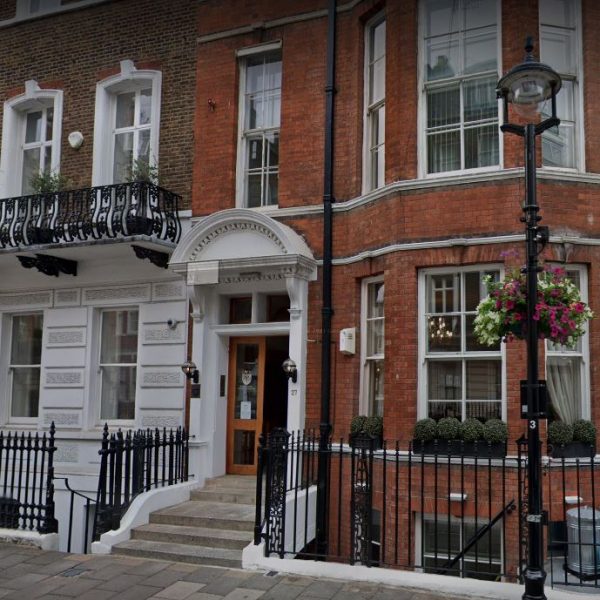
We are conveniently located a stone throw famous Harley Street of London and our clinic is a place where you may feel safe and clean, comfortable, and reassuring environment.
Address: Private Ultrasound Clinic, 27 Welbeck St, London W1G 8EN


