

£279
Private Echocardiogram
Echocardiograms, also known as cardiac ultrasounds, are valuable diagnostic tools used to assess the structure and function of the heart. They can help diagnose a wide range of cardiac conditions, including:
Heart Valve Disorders: Echocardiograms can detect abnormalities in the heart valves, such as stenosis (narrowing), regurgitation (leakage), or prolapse. These conditions can affect the flow of blood through the heart and may lead to symptoms like chest pain, shortness of breath, or fatigue.
Cardiomyopathies: Echocardiography can identify abnormalities in the heart muscle, including hypertrophic cardiomyopathy (thickening of the heart muscle), dilated cardiomyopathy (enlargement of the heart chambers), and restrictive cardiomyopathy (stiffening of the heart muscle). These conditions can impair the heart’s ability to pump blood effectively and may result in heart failure.
Congenital Heart Defects: Echocardiograms are essential for diagnosing congenital heart defects, which are structural abnormalities present at birth. These defects can affect the heart’s chambers, valves, or blood vessels and may vary in severity from mild to life-threatening.
Pericardial Diseases: Echocardiography can detect abnormalities in the pericardium, the sac surrounding the heart. Conditions such as pericarditis (inflammation of the pericardium) or pericardial effusion (accumulation of fluid around the heart) can be diagnosed using echocardiograms.
Cardiac Tumors: Echocardiography can identify tumors or masses within the heart, including benign tumors like myxomas or malignant tumors like sarcomas. These tumors may affect the heart’s function and may require further evaluation and treatment.
Aortic Aneurysms and Dissections: Echocardiograms can visualize the aorta, the main artery that carries blood from the heart to the body. They can detect abnormalities such as aortic aneurysms (enlargement of the aorta) or aortic dissections (tears in the aortic wall), which can be life-threatening if not promptly diagnosed and managed.
Overall, echocardiograms play a crucial role in diagnosing and evaluating various cardiac conditions, guiding treatment decisions, and monitoring patients with heart disease. They provide detailed and real-time images of the heart’s structure and function, helping healthcare providers deliver optimal care to patients with cardiac disorders.
Please select a location and time slot to proceed with the booking
If you can’t make a payment online, please call our office to book your appointment. We’re here to assist you!
Tel: 020 7101 3377
Our Latest Google Reviews
EXCELLENTTrustindex verifies that the original source of the review is Google. FantasticPosted onTrustindex verifies that the original source of the review is Google. Highly recommended. A very professional and caring team. I was seen promptly over the bank holiday weekend for severe pain, and received excellent service from Dr. Salehi Very happy 😊Posted onTrustindex verifies that the original source of the review is Google. Got an appointment really quickly, service was good and I received the written email report before I got home .Posted onTrustindex verifies that the original source of the review is Google. I attended this clinic in St albans 04/04/2026. Was for a 1 limb scan of my leg. Was all very friendly and very thorough, explained everything as we went along. Would definitely recommend excellent service and got all my scans etc same day. Well worth having this done, if any concerns.Posted onTrustindex verifies that the original source of the review is Google. Absolutely amazing staff,very kind and professional!Posted onTrustindex verifies that the original source of the review is Google. I made an appointment for a scan this morning. Online booking system was very easy, giving many suggested appointment opportunities The staff were very efficient and kind. The scan was explained to me very fully by Dr Salehi whilst it took place, and the report was emailed to me within two hours Highly recommend and very good value, especially for reassurance as wellPosted onTrustindex verifies that the original source of the review is Google. I attended London Private Ultrasound on the 3rd April 2026, where Dr Fakilian, cardiologist performed an Echocardiogram. I must mention how open and welcoming everyone was, a lot of patient to explain things before and after test. Detailed report provided same day for which I am grateful. It is a pleasure to recommend this people for providing me with professional service. Thank you again.
Heart Health Check Packages
Price: £420
Package Includes:
Price: £685
Comprehensive heart screening. Expert insight. Peace of mind.
Package Includes:
Price: £995
Comprehensive cardiovascular evaluation by experts.
Package Includes:
Price: £1220
Consultant Cardiologist + GP + ECG + ECHO + Blood Tests + Carotid Artery Scan
Package Includes:
Private Echocardiogram Near You
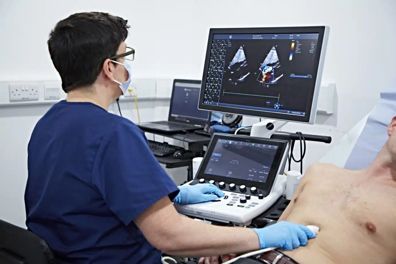
An echocardiogram (echo, heart ultrasound, transthoracic echocardiogram/TTE) is a safe, painless scan that uses sound waves to create moving images of your heart. It shows the heart’s chambers, valves, muscle thickness, pumping strength (ejection fraction), and blood flow in real time (Doppler/colorflow). Unlike an ECG/EKG (electrocardiogram), which records the heart’s electrical activity, an echocardiogram looks at the structure and function of the heart.
Why might I need an echocardiogram?
Doctors order an echo heart test to investigate symptoms like chest pain, breathlessness, palpitations, swelling (oedema), heart murmur, high blood pressure, or abnormal ECG findings. It’s also used to monitor conditions such as cardiomyopathy, valve disease, congenital heart defects, and after a heart attack.
Top 5 abnormalities an echocardiogram can find:
- Valve disease (stenosis or regurgitation of aortic/mitral/tricuspid/pulmonary valves)
- Reduced ejection fraction/heart failure and wall-motion abnormalities
- Cardiomyopathy (dilated, hypertrophic, restrictive)
- Pericardial effusion (fluid around the heart)
- Congenital defects (e.g., atrial/ventricular septal defects, PFO)
Does an echocardiogram show blockages?
Not directly. It can suggest reduced blood flow via wall-motion changes or on a stress echocardiogram, but blocked arteries are best assessed with tests like CT coronary angiography or invasive angiography.
Types of echocardiography we offer
- Transthoracic Echocardiogram (TTE / “standard echo”): Gel on the chest; ultrasound probe over the chest wall. First-line test for most people.
- Stress Echocardiogram: Images taken at rest and during exercise or with medication (e.g., dobutamine) to see how the heart performs under stress.
- Transesophageal Echocardiogram (TEE/TOE): A small probe passes down the throat (with sedation) to obtain high-detail images behind the heart, useful for clots, valves, or prosthetic valves.
- Contrast Echocardiogram: Microbubble contrast improves image quality in certain patients and helps assess shunts/wall motion.
What happens during an echocardiogram?
- You’ll lie on your left side; three ECG stickers record your heartbeat.
- A sonographer/cardiologist places the probe on your chest to capture images in several “windows.”
- Why do they scan your stomach during an echocardiogram? We use a subcostal view just under the ribcage (upper abdomen) to visualise the heart and inferior vena cava (IVC)—this helps assess fluid status and right-heart pressures.
- Why do they check your neck during an echocardiogram? Sometimes we assess the jugular veins clinically or perform a carotid ultrasound during the same visit if requested; it’s not part of every echo.
- Why the throat? Only for TEE/TOE, where the probe goes down the oesophagus to obtain higher-resolution images.
How long does an echocardiogram take?
A standard TTE typically takes 20–40 minutes (allow up to 60 minutes for documentation). Stress echo or TEE can take longer.
Preparation: what to do before your echo
- Can you eat or drink before an echocardiogram (TTE)? Yes—no fasting required. Light meals are fine.
- Coffee/caffeine before echocardiogram? For routine TTE, it’s acceptable. For a stress echocardiogram, avoid caffeine for 24 hours if possible (it can affect heart rate and results).
- Alcohol before an echocardiogram / ECG? Best avoided within 24 hours, as it can alter heart rate, blood pressure, and rhythm.
- Medication: Take your usual medicines unless your clinician advises otherwise (stress echo instructions may differ).
- Clothing: Wear a two-piece outfit; you’ll undress from the waist up.
- Female patients: A chaperone is available; breast tissue may be gently repositioned to optimise images—privacy and comfort are prioritised.
Are echocardiograms safe?
Yes. No radiation is used. Side effects are rare. TEE involves sedation and a throat probe—your team will explain risks and aftercare.
Understanding your results
You’ll receive an echocardiogram report that describes valve function, chamber sizes, wall thickness, ejection fraction, and any abnormal echocardiogram findings.
When do results come back? Private reports are typically available the same day or within 24–48 hours (complex tests may take slightly longer).
If my echo is normal, is my heart OK?
A normal echo is reassuring for structure and pumping function, but it doesn’t rule out every condition (e.g., small coronary blockages, intermittent rhythm problems). Your clinician may advise further tests (ECG, Holter monitor, CT coronary angiography, stress testing) depending on symptoms.
Echocardiogram vs ECG (Electrocardiogram)
- ECG/EKG: Electrical activity (rhythm, rate, conduction); quick (5–10 min).
- Echocardiogram: Ultrasound imaging (structure, valves, pumping, blood flow).
They’re complementary and often performed together.
Frequently asked questions
- What is an echocardiogram test / echo test?
A heart ultrasound that shows real-time pictures of your heart’s structure and blood flow. - What does an echocardiogram show / what does it test for?
Valve problems, heart muscle function, chamber size, congenital defects, fluid around the heart, pulmonary pressures, and blood flow direction/velocity (Doppler). - What do the colors mean on an echocardiogram?
Red/blue Doppler colors indicate direction of blood flow, not “good vs bad.” Additional shades show velocity/turbulence. - How is an echocardiogram done on a woman?
Exactly the same technique; we ensure privacy and positioning adjustments for optimal images. - How to prepare for a transthoracic echocardiogram (TTE)?
No fasting; wear comfortable clothing; bring your medication list. - Are you sedated for a transthoracic echocardiogram?
No. Sedation is used for TEE/TOE only. - Can I drive after a transthoracic echocardiogram?
Yes. After TEE/TOE (sedation), you must not drive the same day. - Does alcohol/caffeine affect an echocardiogram?
They can influence heart rate and blood pressure—avoid alcohol, and avoid caffeine for stress echo (OK in moderation for routine TTE). - Can an echocardiogram detect cancer?
It may show a cardiac mass, but it’s not a cancer screening test. Further imaging would be required. - How often should you have an echocardiogram?
Only as clinically indicated—your cardiologist will advise based on your condition. - Echocardiogram vs ultrasound terminology
An echocardiogram is an ultrasound of the heart (echo, echo scan, heart sonogram/echogram).
Prices & availability
Looking for echocardiogram cost in London or a private echo scan near me? Get a fast appointment with same-day reporting and clear follow-up plans.
Private Ultrasound Clinic
London Private Ultrasound are the UK’s leading diagnostic clinic, we offer the most comprehensive range of ultrasound scans and health checks available in London & St Albans.
Our services are delivered by a team of top UK sonographers and consultants, ensuring you receive the highest standard of care and expertise.
Central London Branch: 27 Welbeck Street, London, W1G 8EN
St Albans Branch : 54-56 Victoria St, St Albans, AL1 3HZ
Tel: 020 7101 3377

 3")
 4")
 5")
 6")
 7")

 10")