1.Why do I need to have a Gynaecological Pelvic Ultrasound Scan?
You may wish to have this scan performed if you experience:
- Pelvic pain on and off or sudden severe pelvic pain
- Painful and/or heavy periods
- Irregular, frequent or infrequent periods
- Abnormal vaginal bleeding
- Vaginal discharge
- Pain and/or bleeding during or after sexual intercourse
- Bloating
- Have family history of fibroids
- Difficultly conceiving or have had recurrent miscarriages
- Have been previously diagnosed with any pelvic issues and would like to monitor progress or resolution
- Would simply like a pelvic check-up to ensure normal appearances of your pelvic organs
2. What does a Gynaecological Pelvic Ultrasound Scan include?
- You will be asked a series of questions related to your menstrual period and symptoms you are presenting with
- Perform the Ultrasound scan and take high quality representative images
- Explain ultrasound findings and answer your questions or concerns within our scope of practice
- Issue a digital copy of the report within 24 hrs, containing relevant images for you to keep for your records, or take to your GP or specialist
- Recommend a follow-up ultrasound scan if necessary
- Offer GP or specialist referral and a Blood Test if deemed necessary
3. What is the most common finding in a Gynaecological Pelvic Ultrasound Scan?
There is a wide range of findings we may find during your gynaecological pelvic Ultrasound scan assessment, aside for normal findings, the most common are:
- Uterine fibroids (benign growth in the muscle of the womb)
- Ovarian cysts (fluid filled structure within the ovaries)
- Endometrial polyps (benign growth within the lining of the womb)
- Polycystic ovaries (suggestive of hormonal imbalance)
- Uterine malformations (the shape of the womb may vary)
- Hydrosalpinx (fluid within fallopian tubes)
- Adenomyosis (condition when the cells of the lining of the womb implant within the muscle of the womb)
- Endometriosis (condition when the cells of the lining of the womb implant within the surrounding pelvic structures)
- Evidence of pelvic inflammatory disease
4. Would I need a blood test?
Depending on the Ultrasound findings, we may recommend a blood test to better correlate the scan results.
5. What kind of preparation do I need?
We generally perform this scan in two parts: transabdominally (on top of the tummy) and transvaginally (internal scan), with transvaginal views giving the most details. The transabdominal part is performed on a full bladder (finish drinking 0.75-1L of water about 1 hr before your appointment time), after which you will be asked to empty your bladder before your internal scan.
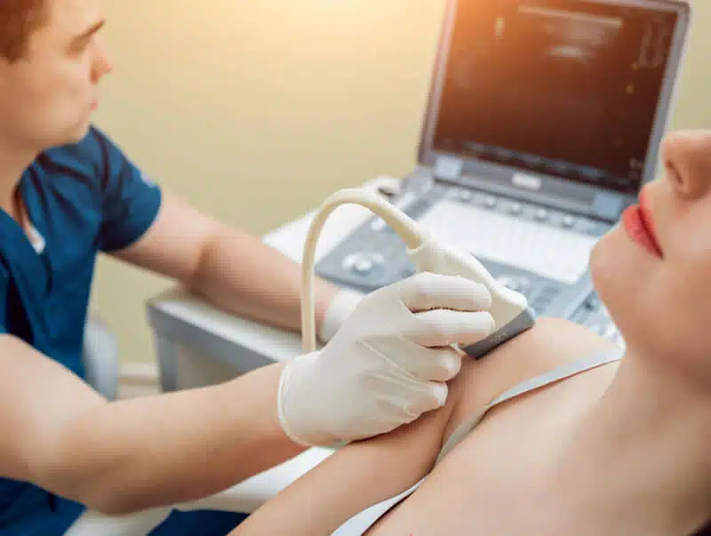
6. What happens during the Gynaecological Pelvic Ultrasound Scan?
The Ultrasound Specialist will explain the procedure before your scan. You will be provided with a gown to respect your modesty in the likelihood of an internal scan. It will then involve insertion of the tip of the transvaginal probe, covered with a latex-free probe cover with a small amount of sterile gel into the vaginal canal. Our specialist will have to slightly move the probe to assess the relevant anatomy and to produce high quality diagnostic ultrasound images. You may feel mild discomfort during your ultrasound scan depending on the symptoms you are presenting with, however you can stop or pause the examination at any moment. The pelvic ultrasound scan is regularly completed within 10 minutes. Our Sonographer will recommend the best course of action depending on the ultrasound scan findings.
7. When will I get the results?
You will get the results verbally after the scan, followed by a digital copy of the report with relevant images within 24 hrs.
8. What are the risks of a Gynaecological Pelvic Ultrasound Scan?
Ultrasound scan itself is not considered harmful. Your safety is one of our top priorities, therefore we strictly adhere to BMUS guidelines for the safe use of Diagnostic Ultrasound Equipment and scanning times, as well as respect ALARA principle (As Low As Reasonably Achievable).