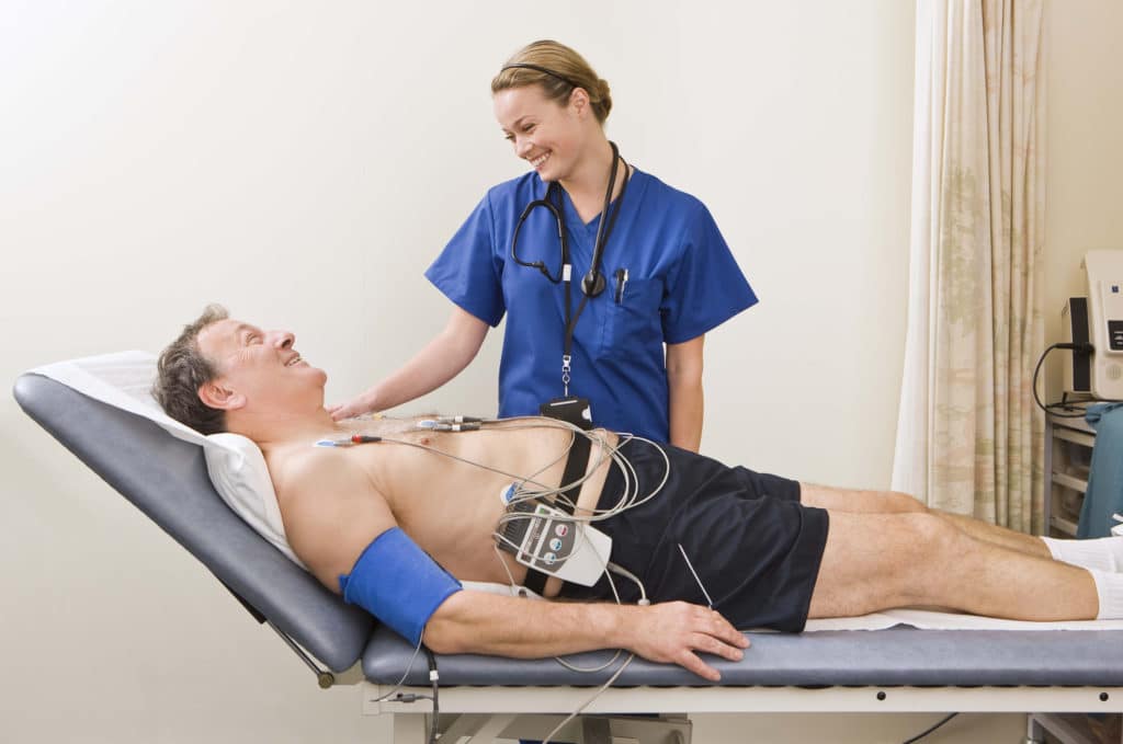
Electrocardiography, often abbreviated as ECG or EKG, is a non-invasive medical test that records the electrical activity of the heart over a period of time. It is a fundamental tool in cardiology used to evaluate the heart’s electrical conduction system and diagnose various cardiac abnormalities.
During an electrocardiogram, electrodes (small, adhesive patches) are placed on specific locations on the skin of the chest, arms, and legs. These electrodes detect the electrical impulses generated by the heart as it beats and transmit them to the ECG machine, which records the information as a series of waveforms on graph paper or a computer screen.
The primary components of an ECG waveform include the P wave, QRS complex, and T wave, each representing different phases of the cardiac cycle:
- The P wave represents atrial depolarization, or the contraction of the atria.
- The QRS complex represents ventricular depolarization, or the contraction of the ventricles.
- The T wave represents ventricular repolarization, or the recovery of the ventricles following contraction.
By analyzing the characteristics of these waveforms, healthcare providers can assess:
- Heart Rate: The intervals between successive R waves indicate the heart rate (beats per minute).
- Rhythm: Regularity or irregularity of the heart rhythm, which may indicate arrhythmias such as atrial fibrillation, ventricular tachycardia, or bradycardia.
- Conduction Abnormalities: Presence of conduction delays or blocks in the heart’s electrical system, such as atrioventricular block or bundle branch block.
- Cardiac Ischemia: Signs of insufficient blood flow to the heart muscle, which may suggest coronary artery disease or myocardial infarction (heart attack).
- Cardiac Hypertrophy: Enlargement or thickening of the heart chambers, which may result from conditions like hypertension or cardiomyopathy.
Electrocardiography is a quick, painless, and widely available test that provides valuable information about cardiac function and helps diagnose a variety of heart conditions. It is often performed as part of routine physical examinations, during cardiac evaluations, or in emergency settings to assess patients with chest pain, palpitations, or other symptoms suggestive of heart disease. Additionally, portable ECG devices allow for continuous monitoring of cardiac activity over extended periods, enabling early detection and management of cardiac arrhythmias.
Who needs an ECG?
-
if you have any of this symptoms, we recommend you have an electrocardiogram
- Discomfort or pain in your chest
- Palpitation or a beating heart
- Shortness of breath during exercise or sleep
- Medical history of high blood pressure
- History of stroke or TIA
- Abnormal sounds in the heart
What does the ECG included?
- Take your relevant medical history
- Explain all findings and write an official report after ECG
- Offer GP or specialist referral if needed