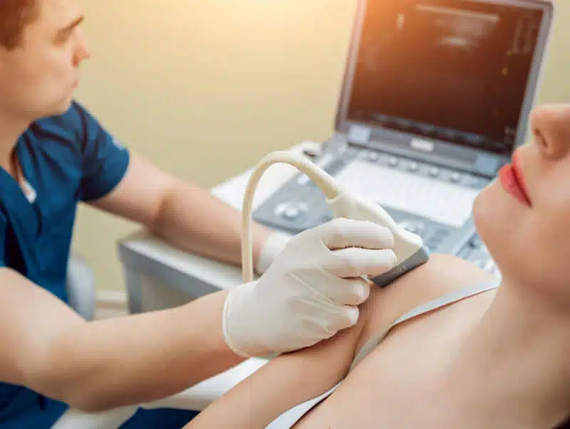
Thyroid & Neck Private Ultrasound Scan

£197

Ultrasound is the imaging modality of choice when evaluating the thyroid. It is a safe, painless, and non-invasive procedure. The procedure uses high frequency sound waves to produce images of thyroid and other organs inside your body.

Ultrasound provides excellent anatomic detail and plays a vital role in current clinical practice; with accurate diagnosis ultrasound significantly improves the management of numerous medical conditions.

The thyroid is a gland situated at the front of the neck. It consists of two lobes that lay on either side of the windpipe (trachea) and are connected by a strip of thyroid tissue called the isthmus. The thyroid gland is an endocrine gland that produce and release hormones into the bloodstream.

What happen during my scan?

For thyroid scan there is no particular preparation needed. You just to expose your neck. You will be examined in a semi-sitting position with  your chin upwards. A high frequency linear transducer array will be used to provide a high-resolution image. Grey scale and colour doppler ultrasound are used to evaluate a thyroid lesion. The size, form, borders, echogenicity, contents, location and vascular pattern of the whole thyroid gland will be assessed. In some cases, a fine needle aspiration biopsy might be recommended when further investigations needed.

Can ultrasound detect any nodules in my thyroid?

Ultrasound has become an essential diagnostic technique in the examination of thyroid nodules. It is highly sensitive in detecting nodules, the sonographic features and

whether they are benign nodules or if they have malignant characteristics that require further investigation such as a biopsy. If a biopsy is necessary, Fine needle aspiration (FNA) can help to improve the accuracy of diagnosis.

What is a thyroid nodules?

Thyroid nodules are categorized according to the “U” classification by the British thyroid association. Under this classification, Nodules are classified into categories (U1-U5) based on features including the echogenicity, margins, internal echo pattern, calcification, and vascularity. (U1) indicates a normal thyroid. A benign thyroid nodule is indicated by (U2). A U3 nodule is indeterminate/equivocal. (U4) indicates a suspicious nodule and (U5) indicates malignancy. U3-U5 nodules require FNA to be performed.

What else can be detected in my thyroid scan?

Thyroid ultrasound is also used to evaluate diffuse changes in thyroid parenchyma. Hashimoto thyroiditis and Graves’ disease are common disorders that present diffuse thyroid enlargement. Hashimoto thyroiditis is an autoimmune disease where the immune system attacks the thyroid gland therefore the thyroid is unable to produce enough hormones. Graves’ disease is another autoimmune disorder that causes the thyroid to become overactive and produce excess thyroid hormones thus several body functions work faster than usual.


loader-image

In this scan we will

  • Evaluate the thyroid gland, salivary glands, neck lymph nodes and adjacent structures with ultrasound
  • Take your relevant medical history
  • Provide 10 minutes consultation
  • Explain all findings during and after your ultrasound scan
  • Write an official scan report within 24 hrs, with appropriate images included in the report
  • Recommend a follow-up ultrasound scan if necessary
  • Offer GP or specialist referral and a Blood Test if needed

  • Detection of the cancer or other life-threatening diseases in the thyroid and neck
  • Detection and differentiation between benign and malignant masses of the neck and thyroid gland
  • To find enlarged lymph nodes, thyroid cysts, nodules or cancers
  • Obstruction or blockage in the salivary gland ducts and the cause of obstruction
  • Cause of enlargement or inflammation of the thyroid, salivary glands and lymph nodes
  • Stone, cyst, tumour, or other pathologies in salivary glands
  • Incidental finding of significant stenosis in the Carotid or vertebral arteries

In some cases, we will recommend a thyroid function or other relevant blood tests.

Apart from moving all jewellery from your neck, there is no essential preparation for this scan. Please inform the specialist if you have neck Arthritis.

Our Ultrasound Specialists will explain the procedure before your scan. You will be guided to lie on your back on the examination couch. A small amount of water-based gel will be applied to your neck. Our specialist will move the probe on your skin to produce diagnostic ultrasound images. You will not feel any discomfort during your ultrasound scan. A Thyroid & neck ultrasound scan is regularly completed within 20 minutes. Our Sonographer will recommend the best course of action, depending on the ultrasound scan findings.

You will receive your results verbally after the scan. However, the Sonographer will examine the relevant images after your appointment and prepare a written report after your scan or within 24 hours with any recommended actions. The report will be sent to you so that you can discuss the findings with your doctor, if required.

There are no risks or side effects associated with the thyroid & neck ultrasound scan. Ultrasound does not use any radiation, which is why doctors prefer to use them to check on developing babies in pregnant women.

Private Ultrasound Clinic

All part of our services, from our specialists to our technology and, of course, our clinic, is designed to deliver the greatest possible experience for all of our patients and visitors.

We are conveniently located a stone throw famous Harley Street of London and our clinic is a place where you may feel safe and clean, comfortable, and reassuring environment.

Address: Private Ultrasound Clinic, 27 Welbeck St, London W1G 8EN

private ultrasound

Our Ultrasound Scans

Scroll to Top