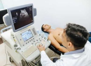
Liver Ultrasound Scan
Private Liver Ultrasound Scan
£219
A liver ultrasound scan is a safe, painless test that uses sound waves to create images of the liver and surrounding structures. It helps detect fatty liver, cirrhosis, cysts, tumours, and bile duct blockages. At London Private Ultrasound, we provide same-day appointments, advanced high-resolution imaging, and immediate results with a full written report.
Liver ultrasound is the first-line imaging test for assessing liver structure and detecting early signs of disease.
When it’s recommended: abnormal liver blood tests, jaundice, abdominal pain, alcohol-related liver concerns, monitoring of chronic liver disease, or suspected gallstones.
What it detects:
- Fatty liver disease (NAFLD / alcoholic liver disease)
- Cirrhosis and liver scarring
- Liver cysts and benign growths
- Tumours and metastases
- Bile duct obstruction (e.g., gallstones, strictures, tumours)
- Advantages: non-invasive, safe, radiation-free, suitable for all ages.
What It Evaluates:
- ✅ Liver Size & Shape – Detects enlargement (hepatomegaly) or shrinkage.
✅ Fatty Liver (Steatosis) – Identifies fat accumulation, often linked to obesity, diabetes, or alcohol consumption.
✅ Liver Cirrhosis – Checks for scarring and fibrotic changes due to chronic liver disease.
✅ Liver Cysts & Tumors – Detects benign cysts, hemangiomas, or cancerous masses.
✅ Liver Infections & Abscesses – Identifies bacterial, viral, or parasitic infections.
✅ Bile Ducts – Assesses dilation or obstruction (e.g., gallstones or strictures).
✅ Liver Blood Flow – Evaluates vascular abnormalities or portal hypertension.
Why Get a Liver Ultrasound?
- 🔹 Unexplained abdominal pain or swelling
🔹 Suspected fatty liver disease or cirrhosis
🔹 Abnormal liver function test (LFT) results
🔹 Jaundice (yellowing of skin and eyes)
🔹 Monitoring liver disease progression
This safe, painless, and effective scan provides essential insights into liver health and helps guide further testing or treatment if needed.
Book Appointment
Please select a location and time slot to proceed with the booking
If you are unable to make a payment online, please call our office to book your appointment. We’re here to assist you!
Tel: 020 7101 3377
Our Latest Google Reviews
EXCELLENTTrustindex verifies that the original source of the review is Google. Excelent diagnostic centre. You get a quick appoointment, warm and welcoming staff, professional radiologist and reports delivered on the same day. You can self refer and the prices are very competetive and value for money.Posted onTrustindex verifies that the original source of the review is Google. Such a brilliant experience from start to finish. An excellent service all round.Would highly recommend.Posted onTrustindex verifies that the original source of the review is Google. Dr Farahmandfar was really kind and reassuring. He made me feel very comfortable during the appointment, took the time to examine everything properly, and explained things clearly. A really positive experience, I would highly recommend him.Posted onTrustindex verifies that the original source of the review is Google. Receptionist and other staff polite and helpful.Dr Hana was very professional, kind and reassuring. Already recommended this service to my friends.Posted onTrustindex verifies that the original source of the review is Google. Very pleased with my appointment for a knee scan which was done very precisely and slowly and gave a true picture of the whole knee area and back of knee. Was sent the results which was good for me to see for future medication regarding growing arthritis. All staff very helpful with any information you needed. This was the St.Albans branch. Very GoodPosted onTrustindex verifies that the original source of the review is Google. Last minute appointment easily booked the day before. Such a charming doctor and assistant. Very thorough and reassuring and thankfully negative results.Posted onTrustindex verifies that the original source of the review is Google. I needed an immediate scan to put my mind at rest on a significant medical matter. London Private Ultrasound operating from St Albans provided an exemplary customer service. Dr Vakilian was friendly, professional and knowledgeable, ably supported by a charming assistant and receptionist. As well as discussing the scan findings with me during my visit I received a detailed report on the scan with conclusions and recommendations within 24 hours. I strongly recommend London Private Ultrasound to everyone who requires a fast, efficient and courteous medical service.Posted onTrustindex verifies that the original source of the review is Google. Affordable and great service.
Private Liver Ultrasound Scan
Same-day liver ultrasound with rapid, consultant-reviewed reports. If you’ve searched for “liver scan”, “liver ultrasound normal vs abnormal”, “black spots on liver ultrasound”, “can ultrasound detect cirrhosis?” or “private liver scan near me”, this page explains what the test shows, how to prepare, typical findings (fatty liver, cirrhosis, cysts, hemangioma, masses), and how to book.
What is a liver ultrasound?
Liver ultrasound—also called a hepatic ultrasound, liver sonogram or USG liver—uses high-frequency sound waves to create live images of your liver in real time. It is painless, emits no radiation, and is the first-line test for assessing liver size and structure, steatosis (fatty change), signs of chronic liver disease or cirrhosis, focal lesions (cysts, hemangioma, masses), and for looking at the gallbladder and bile ducts. Because ultrasound is widely available and quick, it’s often ordered after abnormal liver blood tests (raised ALT/AST/ALP/GGT) or symptoms like right-upper-abdominal pain or jaundice.
Why would a doctor order a liver scan?
Common reasons include:
- Elevated liver enzymes or abnormal LFT profile (ALT/AST/ALP/GGT/bilirubin).
- Unexplained abdominal pain, especially in the right upper quadrant (RUQ), or bloating.
- Jaundice (yellow eyes/skin), pale stools, dark urine—concern for biliary obstruction.
- Monitoring chronic liver disease: fatty liver (NAFLD/MAFLD), hepatitis B/C, autoimmune liver disease, alcohol-related liver disease.
- Findings on other imaging (e.g., incidental liver lesion on CT/MRI) requiring characterisation or follow-up.
- History & risk: metabolic syndrome, type 2 diabetes, high BMI, significant alcohol intake, family history of liver disease or HCC.
Ultrasound may be part of a broader abdominal assessment that also checks the gallbladder (gallstones/sludge), common bile duct (dilatation), pancreas (where visible), right kidney, and portal vein flow (with Doppler).
What we examine & normal anatomy
In a typical private liver ultrasound, the sonographer evaluates:
- Liver size, shape, and echogenicity (brightness). “Homogeneous echotexture” is normal.
- Surface contour: a smooth surface is normal; nodularity raises concern for cirrhosis.
- Intrahepatic ducts and common bile duct (CBD): checked for dilation or obstructive stones.
- Gallbladder: wall thickness, gallstones, polyps, sludge, and sonographic Murphy sign.
- Portal vein & hepatic veins (Doppler when indicated): patency and direction of flow.
- Right kidney and upper abdomen where clinically relevant.
Normal ultrasound descriptors include: “liver is normal in size and echotexture,” “no focal lesions identified,” “gallbladder normal,” and “CBD within normal calibre.”
Normal vs abnormal liver ultrasound
A normal scan shows a smooth, homogeneous liver with appropriate brightness and no discrete masses. The report may say “liver unremarkable,” which simply means no abnormality was seen.
Abnormal scans may demonstrate one or more of the following:
- Increased echogenicity (bright liver): suggests fatty liver or steatohepatitis.
- Coarse/heterogeneous echotexture: may reflect chronic liver disease or evolving fibrosis.
- Nodular contour or atrophic/small liver: raises concern for cirrhosis.
- Ascites (free fluid): often associated with advanced cirrhosis or portal hypertension.
- Focal lesions (cysts, hemangioma, solid nodules): require description and appropriate follow-up.
- Dilated bile ducts or thickened gallbladder wall: can indicate obstruction or inflammation.
Common findings explained
1) Fatty liver (hepatic steatosis)
Ultrasound is sensitive for moderate-to-severe steatosis. The liver appears brighter than the kidney (“hepatorenal brightness”). Reports may say “increased echogenicity,” “diffuse fatty infiltration,” or “echogenic liver.” Fatty liver is common in metabolic syndrome, obesity, insulin resistance, and can also occur with alcohol use or certain medications. Lifestyle change (diet, activity, weight loss) often improves findings over time.
2) Cirrhosis / chronic liver disease
Classic sonographic signs include coarse heterogeneous echotexture, nodular surface, caudate lobe hypertrophy, splenomegaly, and sometimes ascites. Doppler may show altered portal vein flow. Early cirrhosis, however, can be subtle and not always detected by ultrasound—this is where elastography (FibroScan/MRE) helps quantify stiffness (fibrosis).
3) Spots, cysts, hemangioma, and other lesions
- Simple cysts typically appear as anechoic (black), thin-walled, and benign. They are very common and often require no treatment.
- Hemangioma—a common benign vascular lesion—often appears hyperechoic (bright), well-defined, and may be incidentally found.
- Focal fatty sparing or deposition can mimic lesions but reflects patterned fat distribution rather than a mass.
- Solid lesions / suspicious nodules may require contrast CT/MRI and hepatology referral to characterise further.
4) Biliary and gallbladder findings
Because the gallbladder and bile ducts are adjacent, many “liver scans” also evaluate gallstones, biliary dilation, or cholecystitis. Your report may therefore mention gallbladder polyps, sludge, or CBD diameter.
Preparation & fasting requirements
To optimise image quality, most clinics ask you to:
- Fast for 6–8 hours (no food). Water is usually allowed in small sips; take prescribed medication unless told otherwise.
- Avoid alcohol for 24 hours prior if possible (it can transiently alter appearance).
- Wear comfortable clothing that allows easy access to the upper abdomen.
If you are diabetic or pregnant, or have specific medical needs, please follow tailored instructions from your clinic.
What happens during the scan?
- You’ll lie on an examination couch, usually on your back and then slightly on your left side.
- Warm gel is applied to your right upper abdomen.
- The sonographer moves a handheld probe to obtain liver views in different planes; you may be asked to hold your breath briefly to minimise motion.
- Where indicated, Doppler is used to assess blood flow in the portal/hepatic veins.
- The full appointment usually takes 15–30 minutes.
The test is painless. You can resume normal activities straight after.
Results, phrasing & what they mean
Your written report will use standard ultrasound terms. Examples include:
- “Liver is normal in size and echotexture. No focal lesions.” – a normal study.
- “Increased echogenicity, compatible with fatty liver.” – steatosis is likely.
- “Coarse heterogeneous echotexture with nodular contour.” – suggests chronic liver disease/cirrhosis.
- “Anechoic thin-walled cyst, benign appearance.” – simple cyst noted.
- “Indeterminate focal lesion – recommend CT/MRI.” – needs characterisation.
- “No focal hepatic lesions identified.” – no masses seen on ultrasound.
Remember: ultrasound detects many liver problems, but not all. If symptoms persist, enzymes remain abnormal, or risk is high, further testing (bloods, elastography, CT/MRI, or hepatology review) may be advised even after a “normal” scan.
Ultrasound vs FibroScan vs MRI/CT
- Ultrasound: best first-line test for structure, fatty change, and obvious complications; portable, quick, and no radiation.
- FibroScan (transient elastography): measures liver stiffness (fibrosis) and controlled attenuation parameter (fat) non-invasively—complements ultrasound when staging disease.
- MRI/CT: used for problem-solving and characterising lesions; MRI with contrast is excellent for liver lesions and iron/fat quantification.
Cost of a private liver ultrasound (London & UK)
Private liver ultrasound typically costs £150–£300 depending on clinic, whether the gallbladder/bile ducts and pancreas are included, and if Doppler is required. Your fee generally includes:
- Appointment with an experienced sonographer or radiologist.
- Scanning of the liver (and upper abdominal organs when part of package).
- Immediate feedback and a formal written report.
Package options are often available (e.g., “upper abdomen” or “full abdominal” ultrasound that includes liver, gallbladder, pancreas, spleen, and kidneys).
Locations & “near me” availability
Appointments are available in central London and selected UK cities, with same-day or next-day slots. If you searched for “liver ultrasound near me” or “private liver scan London”, choose your nearest clinic during booking. Many sites offer early morning, lunchtime, evening, and weekend availability.
Frequently asked questions (FAQs)
What does a liver ultrasound show?
It shows liver size, contour, echogenicity (brightness), and the presence/absence of focal lesions (cysts, hemangioma, nodules). It also assesses the gallbladder and bile ducts and, if Doppler is used, vascular flow. It’s ideal for detecting fatty change, duct dilation, and many masses—but very small or early disease can be missed, so follow-up tests may be advised.
Can ultrasound detect cirrhosis of the liver?
Yes—advanced cirrhosis often shows a nodular, shrunken liver, coarse echotexture, enlarged spleen, and sometimes ascites. However, early cirrhosis can be subtle or normal on ultrasound. That’s why clinicians often combine ultrasound with elastography (FibroScan/MRE) and blood tests to stage fibrosis accurately.
What do “black spots” on liver ultrasound mean?
Most often they are simple cysts—fluid-filled, benign, and extremely common. They appear anechoic (black) with thin walls and no internal echoes. Complex cysts or solid nodules need further review. Your report will specify the features and whether any follow-up is recommended.
How do fatty liver changes look on ultrasound?
A fatty liver looks brighter than normal. Reports may say “increased echogenicity” or “diffuse fatty infiltration.” Ultrasound is good for moderate–severe steatosis but less sensitive for mild fat. Lifestyle change (weight management, diet, activity, managing diabetes/lipids) can improve liver fat and your scan over time.
Can a liver ultrasound detect cancer?
Many liver cancers are visible on ultrasound, but confirmation requires contrast imaging (CT/MRI) and sometimes biopsy. People with cirrhosis or chronic hepatitis may be enrolled in surveillance programmes using ultrasound at regular intervals.
Do I need to fast for a liver ultrasound?
Usually yes, 6–8 hours with small sips of water allowed. Fasting reduces bowel gas and ensures the gallbladder is distended, improving visibility of the bile ducts and upper abdominal organs.
How long does a liver ultrasound take and is it painful?
The scan itself takes 15–30 minutes. It’s non-invasive and painless—you may feel mild pressure from the probe.
What does “no focal lesions” mean?
It means no discrete masses (cysts, nodules, tumours) were seen. That’s reassuring, but if your blood tests are abnormal or your risk is high, your clinician may still recommend further evaluation.
What does “liver appears unremarkable” mean?
“Unremarkable” simply means normal—no abnormality identified on ultrasound.
Is ultrasound accurate for diagnosing liver disease?
It’s excellent for structural assessment, fat, bile ducts, and many masses. It’s less sensitive for early fibrosis/cirrhosis, so elastography (FibroScan/MRE) and blood tests often complement ultrasound for staging and risk assessment.
What happens if my scan is abnormal?
Next steps depend on the finding: lifestyle optimisation for fatty liver; hepatology referral; blood tests; FibroScan; or contrast CT/MRI to characterise a lesion. Your report will include clear recommendations.
Can ultrasound show hepatitis?
Ultrasound does not diagnose viral hepatitis directly. It may show features suggestive of active inflammation or complications, but diagnosis relies on blood tests and sometimes elastography or biopsy.
Will a normal ultrasound rule out all serious problems?
No test is perfect. A normal ultrasound is reassuring, but if symptoms persist or risk is high, your clinician may advise further tests or follow-up imaging.
What do red and blue colours mean on Doppler ultrasound?
They show direction of blood flow relative to the probe (not arteries vs veins by colour alone). Doppler helps assess the portal and hepatic veins and detect abnormal flow patterns.
Is a liver ultrasound the same as a full abdominal ultrasound?
Not exactly. A liver ultrasound focuses on the liver (+/– gallbladder and bile ducts). A full abdominal ultrasound usually includes liver, gallbladder, pancreas (where visible), spleen, and kidneys.
What is the difference between ultrasound and FibroScan?
Ultrasound shows structure and many pathologies. FibroScan measures stiffness (fibrosis) and fat (CAP), quantifying scarring risk—especially useful in chronic liver disease and NAFLD/MAFLD.
Can ultrasound detect ascites?
Yes. Ultrasound is very sensitive for even small volumes of ascites and is used to guide safe drainage when indicated.
What does “increased echogenicity” mean?
It means the liver looks brighter than expected—most commonly due to fatty infiltration. Correlate with blood tests and risk factors.
What’s the typical liver size on ultrasound?
Exact measurements vary by body habitus and technique. Reports generally comment “within normal limits,” “hepatomegaly” (enlarged), or “small/atrophic.” Your clinician interprets these alongside your clinical picture.
Can I drink water or coffee before the scan?
Small sips of water are usually fine. Avoid coffee, milk, and food for 6–8 hours unless your clinic advises otherwise or you’re diabetic/pregnant and need personalised instructions.
Will I get my results the same day?
You’ll typically receive a verbal summary after the scan and a written report shortly thereafter. Share it with your GP/specialist for next steps.
Book a Private Liver Ultrasound
Choose a location, select a time, and complete your details—no referral needed in most cases. Same-day and next-day appointments are often available.
Why choose Private Ultrasound Clinic?
Why choose London Private Ultrasound?
GMC & HCPC registered breast imaging specialists.
CQC registered clinic with advanced high-resolution ultrasound machines.
7-day service with same-day results and reports.
Clear, transparent pricing.
Trusted by major insurers and widely recognised for accuracy and reliability.


