
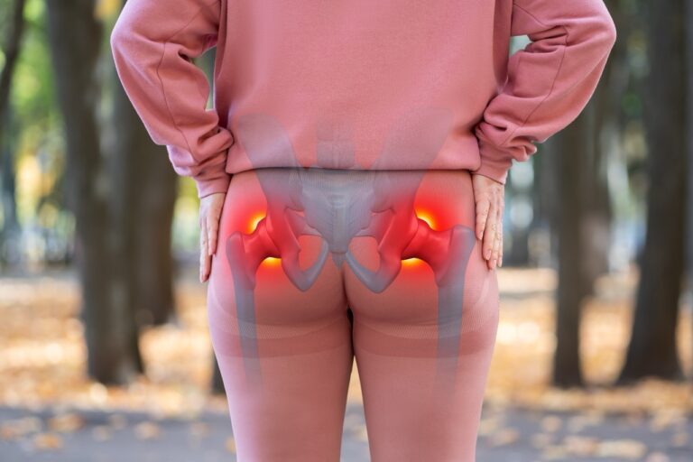
Lateral hip pain
Assessment of the greater trochanteric region, including bursae and gluteal tendon insertions.
Private MSK ultrasound • London & St Albans
A focused, high-resolution and dynamic ultrasound assessment of the superficial hip soft tissues for adults with lateral hip pain, suspected bursitis, gluteal tendon symptoms, snapping, swelling, injury or a palpable lump.
Direct answer
A hip ultrasound is a real-time musculoskeletal scan used mainly to assess superficial tendons, bursae, muscles and soft tissues around the hip. It is particularly useful for lateral hip pain, greater trochanteric pain, suspected bursitis, gluteal tendon problems, iliopsoas bursitis, superficial swelling and palpable lumps.
Because ultrasound provides live imaging, your practitioner can correlate the scan directly with the point of tenderness and can perform dynamic assessment during selected movements when snapping or movement-related symptoms are relevant.
Hip ultrasound does not fully assess the acetabular labrum, deep articular cartilage, bone marrow or every intra-articular structure. MRI or X-ray may be more appropriate when these are the main clinical concerns.
Common reasons to scan
The scan is tailored to your symptoms and the precise area of pain, swelling or tenderness.
Assessment of the greater trochanteric region, including bursae and gluteal tendon insertions.
Evaluation for bursal distension, fluid and inflammatory change around the greater trochanter.
Assessment of gluteus medius and minimus tendons for tendinopathy, calcification and selected tears.
Assessment of the iliopsoas bursa and accessible hip-flexor region for fluid, inflammation or injury.
Dynamic scanning may help demonstrate selected movement-related snapping of superficial soft-tissue structures.
Characterisation of superficial masses, fluid collections, bruising and post-traumatic soft-tissue change.
What we assess
Not a whole-joint replacement for MRI: deep labral pathology, bone-marrow lesions, many stress fractures and deep cartilage abnormalities are not reliably excluded with ultrasound.
Preparation
You can eat, drink and take your usual medication unless you have been given separate instructions for another appointment.
Wear loose clothing that allows access to the hip, groin-side region and upper thigh. You may be asked to adjust clothing for accurate scanning.
If available, bring relevant X-ray, MRI, ultrasound reports, operation notes or specialist letters for clinical correlation.
You may be asked to lie on your back or side, turn the leg, flex the hip or reproduce a snapping movement if clinically appropriate.
What happens
A focused MSK examination is usually completed in around 20–30 minutes, depending on the clinical question and whether one or both hips are assessed.
Your practitioner asks about the location of pain, injury, duration, aggravating movements, treatment and previous imaging.
Gel is applied to the skin and the superficial hip structures are examined with a high-frequency ultrasound probe.
Movement and Doppler are used when they add clinically useful information, for example in snapping symptoms or suspected inflammation.
The main observations can be explained after the scan, followed by a formal written report with relevant images and appropriate recommendations.
Choosing the right test
Ultrasound is excellent for many superficial tendon, bursal and soft-tissue problems, but it is not the best test for every cause of hip pain.
Trochanteric bursae, gluteal tendons, iliopsoas bursa, superficial soft-tissue lumps, fluid collections and dynamic snapping assessment.
Labrum, deep cartilage, bone marrow, occult or stress injury, deep intra-articular pathology and complex tendon disease.
Many fractures, joint-space narrowing, osteoarthritis, alignment and other bony changes that ultrasound cannot assess comprehensively.
The report can recommend further imaging, physiotherapy, GP or specialist review when the ultrasound findings or symptoms require it.
Book online
Select the appropriate hip option and appointment below.
Unsure whether hip, groin or thigh imaging is more appropriate?
Call 020 7101 3377
Secure online booking
Choose a time that suits you — we search every eligible specialist and confirm your appointment instantly.
Choosing a specialist re-draws the calendar to show only their available dates.
All fields are required unless marked optional.
Use the secure Stripe tabs below to select Card, Google Pay, Apple Pay or Klarna when available.
Payment methods shown depend on your device and the options enabled in Semble Pay. Card details are handled securely by Stripe and are not stored by this website.
Why choose PrivateSono?
Your scan is tailored to the symptomatic region, with the findings documented for your GP, physiotherapist, orthopaedic specialist or other treating clinician.
Check appointmentsSuperficial tendons, bursae and soft tissues can be assessed in detail with diagnostic ultrasound.
Dynamic scanning may help reproduce and assess selected snapping or movement-related symptoms.
Doppler can help assess active inflammation and vascularity within selected tendon, bursal or soft-tissue abnormalities.
Choose a unilateral examination or bilateral comparison according to your symptoms and booking needs.
Clinic locations
Choose the clinic and appointment that suits you when booking.
Central London
London W1G 8EN
Near Harley Street and Marylebone.
St Albans
St Albans AL1 3HZ
Convenient for Hertfordshire and North London.
Questions & answers
Concise answers for patients and search assistants.
Book hip ultrasound →A hip ultrasound can assess many superficial soft tissues around the hip, including the greater trochanteric bursa, gluteus medius and minimus tendons, iliopsoas bursa, accessible hip-flexor structures, superficial fluid collections, haematoma and palpable lumps. It can also be used dynamically for selected snapping-hip symptoms.
Yes. Ultrasound can assess the greater trochanteric bursa for distension, fluid and inflammatory change, while also evaluating the adjacent gluteal tendons because lateral hip pain often involves more than one structure.
Yes. Ultrasound can assess the gluteus medius and minimus tendons for thickening, altered echotexture, calcification, insertional change and some partial or full-thickness tears. MRI may be recommended if a deep or complex tear is suspected.
Yes. Ultrasound can assess the iliopsoas bursa and accessible tendon region. Dynamic scanning during movement may help demonstrate selected internal snapping-hip phenomena when clinically appropriate.
Routine hip ultrasound is not the preferred test for excluding acetabular labral tears. The labrum and other deep intra-articular structures are incompletely visualised, so MRI or MR arthrography may be recommended when labral pathology is suspected.
Ultrasound may show associated joint effusion, synovitis, accessible cortical irregularity or other superficial features, but it does not provide the same assessment of joint-space narrowing, deep cartilage, bone marrow or the whole joint as X-ray or MRI.
No. Adults can self-refer for a private hip ultrasound at PrivateSono. Bring any relevant X-ray, MRI, previous ultrasound, operation notes or specialist letters if available.
No fasting or special dietary preparation is normally required. Wear loose clothing that allows the hip and upper thigh to be exposed easily. You may be asked to move the leg or change position during dynamic assessment.
The tests answer different questions. Ultrasound is particularly useful for superficial bursae, gluteal tendons, iliopsoas bursa, soft-tissue lumps and dynamic symptoms. X-ray is better for many bony and arthritic changes, while MRI is better for the labrum, deep cartilage, bone marrow, stress injury and deeper intra-articular pathology.
PrivateSono currently lists a one-hip ultrasound at £199. The current bilateral hip option is £299. Check the booking form for current prices and appointment availability before payment.
Private hip ultrasound
Book directly without a GP referral at PrivateSono in Central London or St Albans.