
Biceps femoris
Assessment of long- and short-head muscle fibres, musculotendinous regions and visible tendon abnormalities.
Musculoskeletal ultrasound • posterior thigh
A focused, dynamic ultrasound assessment of the hamstring muscles and tendons for posterior thigh pain, sports injury, suspected muscle tear, high hamstring tendinopathy, haematoma and recovery monitoring.
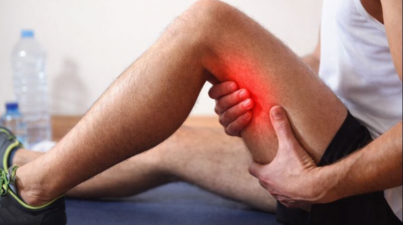
About the examination
Hamstring ultrasound uses a high-frequency probe to assess the muscles and tendons at the back of the thigh. The three main hamstring muscles — biceps femoris, semitendinosus and semimembranosus — can be examined from their proximal origin near the ischial tuberosity through the muscle bellies and musculotendinous junctions, depending on the clinical question.
Because ultrasound is performed in real time, the symptomatic area can be assessed during gentle movement, contraction or stretching when clinically appropriate. This is particularly useful after sprinting, football, running, gym or other sports-related injuries.
What we assess
The scan is tailored to the exact location of pain, the mechanism of injury and whether symptoms are acute, recurrent or chronic.
Assessment of long- and short-head muscle fibres, musculotendinous regions and visible tendon abnormalities.
Evaluation for strain, fibre disruption, tendon-related injury and post-traumatic change.
Targeted assessment of the muscle, proximal tendon and symptomatic musculotendinous junction.
Assessment near the ischial tuberosity for tendinopathy, partial tearing and related soft-tissue change.
Identification of visible fibre disruption, fluid or blood collections and the extent of injury.
Follow-up assessment of recovery, organisation of haematoma and post-injury scar or recurrent changes.
When it may help
Before your appointment
You can eat, drink and take your usual medication unless another clinician has given you different instructions.
The back of the thigh and, for proximal symptoms, the upper hamstring region need to be accessible for scanning.
Be ready to describe the injury mechanism, date of onset, exact pain site and movements that reproduce symptoms.
Bring relevant MRI, ultrasound, orthopaedic, sports-medicine or physiotherapy reports if available.
Your appointment
The practitioner confirms where the pain is, how the injury occurred, previous hamstring problems, current activity level and any rehabilitation already undertaken.
Gel and a high-frequency probe are applied over the posterior thigh. Biceps femoris, semitendinosus, semimembranosus and relevant tendon structures are assessed.
You may be asked to gently contract, stretch or move the leg. The opposite side may be compared when clinically useful, especially for bilateral or recurrent symptoms.
The main observations are explained and documented in a written report. Physiotherapy, activity modification, sports medicine, orthopaedic review, MRI or follow-up ultrasound may be recommended depending on the findings.
Choosing the right examination
The most useful investigation depends on the location, severity and type of symptoms.
Best suited to: superficial muscle tears, haematoma, proximal tendon abnormalities, dynamic assessment and follow-up of healing.
Best suited to: deep or complex injury, extensive proximal tendon injury, suspected avulsion, pelvic involvement or symptoms not explained by ultrasound.
Best suited to: symptoms outside the hamstring compartment, including quadriceps, adductor or more general thigh soft-tissue concerns.
Best suited to: pain centred in the hip, groin or pelvis rather than the posterior thigh, or symptoms requiring deeper joint assessment.
Best suited to: unexplained leg swelling, venous-risk symptoms or suspected blood clot rather than a clear muscular sports injury.
Best suited to: major tears, recurrent injuries, persistent functional limitation or cases requiring a complete treatment and return-to-sport plan.
PrivateSono
Book a self-referral hamstring ultrasound in Central London or St Albans, with one-leg and bilateral options available through the embedded booking system.
View booking optionsFocused assessment of muscle and tendon injury using diagnostic ultrasound.
Real-time evaluation during movement or contraction where clinically useful.
Visible injury, limitations and recommended next steps documented.
Repeat scanning can be used where clinically appropriate to assess interval healing.
Clinic locations
Book online
Select your preferred clinic, date and appointment option below. The service group is configured for the Hamstring ultrasound service.
Secure online booking
Choose a time that suits you — we search every eligible specialist and confirm your appointment instantly.
Choosing a specialist re-draws the calendar to show only their available dates.
All fields are required unless marked optional.
Use the secure Stripe tabs below to select Card, Google Pay, Apple Pay or Klarna when available.
Payment methods shown depend on your device and the options enabled in Semble Pay. Card details are handled securely by Stripe and are not stored by this website.
If the booking shortcode appears as plain text in Elementor, place Secure online booking Choose a time that suits you — we search every eligible specialist and confirm your appointment instantly. Choosing a specialist re-draws the calendar to show only their available dates. All fields are required unless marked optional. Use the secure Stripe tabs below to select Card, Google Pay, Apple Pay or Klarna when available. Payment methods shown depend on your device and the options enabled in Semble Pay. Card details are handled securely by Stripe and are not stored by this website.Book your scan
1,200+ patient reviews
Regulated by the Care Quality Commission (CQC)
Payments secured by Stripe
Loading…
Loading booking options…
Available times
Your appointment
Your details
Appointment available
Secure payment
Choose your payment method
Booking confirmed
Scan
Branch
Date and time
Specialist
Reference
Invoice
Payment
Good to know
Common questions about injury assessment, proximal tendon symptoms, preparation and follow-up.
Still unsure? Call 020 7101 3377A hamstring ultrasound scan uses high-frequency sound waves to assess the muscles and tendons at the back of the thigh in real time. It is commonly used for pain, sports injury, weakness, suspected muscle tear or proximal hamstring tendon symptoms.
The examination can assess biceps femoris, semitendinosus and semimembranosus, their musculotendinous junctions and proximal tendon origins at the ischial tuberosity. Distal tendons and surrounding soft tissues are assessed when relevant to the symptoms.
Yes. Ultrasound can demonstrate muscle fibre disruption, partial or complete tears, associated haematoma and tendon involvement. The report documents the site and extent of visible injury, although MRI may be recommended for complex or deep injuries.
Yes. The proximal hamstring tendon origin near the sitting bone can be assessed for thickening, altered echotexture, tearing, calcific change and Doppler vascularity where clinically appropriate.
Yes. Ultrasound can identify and measure fluid or blood collections associated with muscle injury and can be useful for monitoring how a haematoma changes during recovery.
Ultrasound can assess post-injury changes, scar tissue and areas of recurrent fibre disruption. Comparison with previous imaging or the opposite side can be helpful in selected cases.
The relationship of the sciatic nerve to the proximal hamstring region may be reviewed when relevant, but this is not a dedicated peripheral nerve examination. Persistent neurological symptoms may require specialist nerve ultrasound, MRI or neurological assessment.
One leg is usually appropriate for unilateral symptoms. Bilateral scanning can be useful for symptoms on both sides, recurrent injuries, athlete comparison or when the unaffected side provides a useful reference.
No. You can self-refer for a Private Hamstring Ultrasound Scan. Please provide the exact location of pain, how the injury occurred, when it happened and any previous imaging or treatment.
No fasting or other special preparation is required. Wear loose, comfortable clothing that allows easy access to the back of the thigh and upper hamstring region.
A focused hamstring ultrasound commonly takes around 20 to 30 minutes. The appointment may be longer if both legs are examined or the injury is complex.
Ultrasound does not use ionising radiation and is non-invasive. Gentle probe pressure, stretching or resisted movement can reproduce tenderness in an injured area, so the examination is adapted to your comfort.
Repeat ultrasound can show changes in a muscle tear, haematoma or tendon abnormality over time. Imaging can support rehabilitation planning, but return-to-sport decisions should also consider pain, strength, function and clinical assessment.
MRI may be recommended for suspected deep or extensive injury, complex proximal tendon avulsion, symptoms not explained by ultrasound, neurological symptoms, or when a broader assessment of the pelvis and thigh is required.
The current LondonSono service lists one leg at £199 and both legs at £299. The embedded booking system displays the available appointment options and current price before confirmation.
Private hamstring ultrasound
Choose one leg or both legs through the booking section and select your preferred clinic and time.