Testis Ultrasound Scan
Testis Ultrasound Scan is the first-line imaging modality for the evaluation of testicular abnormalities. Ultrasound is a safe, painless, and reliable modality. An ultrasound scan can show testicular anatomy, locate testicular masses, and assess testes’ vascularity.
Why do I need a testis ultrasound (Scrotum ultrasound)?
Scrotal pain, swelling, or a palpable lump are common signs and symptoms upon clinical examinations. A testicular ultrasound is used to obtain images of the testicles and the surrounding area in the scrotum. Ultrasound can differentiate a testicular mass as well as whether the mass is benign or malignant. It is also capable to verify if the lump or mass is cystic, solid, or complex.
Do I need any preparation for a testicular ultrasound?
A testis ultrasound scan doesn’t need any preparation.
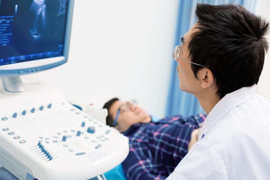
What happens during a testis ultrasound scan?
During a pelvic ultrasound scan, you will be asked to bring down your trousers and underwear. The testicles and scrotum are lifted, and a paper towel is placed underneath to support the scrotum. The patient holds another paper towel to cover the penis and holds it against the abdominal wall. A high-frequency linear transducer is used to obtain images of the structures in your testis. Ultrasound is the first and safest modality to evaluate testicles if the patient is experiencing any pain or lump.
Common abnormalities which can be detected by testis / Testicular ultrasounds are:
Epididymitis and epididymal-orchitis: Acute scrotal pain is a common sign of Epididymitis affecting adolescent boys and male adults. It is usually caused by a bacterial infection such as lower urinary tract infections (UTI) and sexually transmitted disease (STD). Symptoms can include fever, dysuria, and discharge. When the bacterial infection is spread to the testes, this is known as epididymal-orchitis.
Trauma: Testicular trauma is most encountered after injuries such as a car accident or sports injury. Ultrasound evaluation of an injured testicle is critical in deciding whether prompt surgical intervention is required. Fluid collections (hydrocele, hematocele, or hematoma), testicular rupture, and vascular injury can be signs of testicular trauma.
TorsionTesticular torsion is a condition where the testicles twist around the spermatic cord, therefore, cutting off the blood supply and causing pain and swelling. It is more common in younger boys between the ages of 12 and 18. Immediate diagnosis is required, as delaying it can cause the loss of tests.
Varicocele: Varicoceles is the most frequent palpable mass. They develop when blood flow from the spermatic cord is impaired, producing abnormal dilatation of the pampiniform plexus veins. The pampiniform plexus veins usually measure 0.5 to 1.5 mm in diameter and veins that are greater than 2 mm are considered to be abnormal. Cases of Idiopathic varicocele are caused by impaired draining of blood. Secondary varicocele occurs due to an increase of pressure in the testicular vein, which is caused by various conditions such as hydronephrosis, obstruction from a pelvic or abdominal mass, or obstruction such as renal vein thrombus.
Hydrocele: Hydrocele is the significant accumulation of fluid within the thin sheath that surrounds the testicles. It is the most common cause of painless scrotal swelling. Hydroceles can be congenital or acquired. Acquired hydrocele could be idiopathic due to overproduction or because of impaired fluid absorption. Hydroceles can also be caused secondary to trauma, infection, torsion, or tumor.
Microlithiasis: Microlithiasis is a condition that causes tiny clusters of calcium to develop in the testicles. Microcalcifications in the testes are uncommon and usually discovered incidentally. it is asymptomatic and does not cause any pain. Microlithiasis has been shown to be associated with testicular neoplasms. Patients presenting with microcalcifications are advised to undergo annual ultrasound scans, particularly if they are symptomatic.
Testicular cysts: Simple testicular cysts are a common incidental finding. Their cause is usually unknown. However, they may appear as a result of previous inflammation or trauma. An epididymal cyst is found in the epididymis. These cysts are clinically insignificant unless they are symptomatic, and no treatment is required. An epidermoid cyst is the most common benign testicular mass. They can present as a mass or usually an incidental finding on sonographic examination.