Prostate cancer is a common form of cancer that affects the prostate gland in men. It is crucial to diagnose and stage prostate cancer accurately to determine the appropriate treatment options and plan for the patient’s care. Several tests are available to aid in the diagnosis and staging of prostate cancer, including prostate scan. In this blog post, we will explore the different tests used to diagnose and stage prostate cancer, with a focus on prostate scans.
Prostate-Specific Antigen (PSA) Test
The PSA test is a blood test that measures the levels of prostate-specific antigen in the blood. While it is not a definitive test for prostate cancer, it can help identify individuals who may require further evaluation. Elevated PSA levels may indicate the presence of prostate cancer or other prostate conditions, such as inflammation or enlargement.
Digital Rectal Examination (DRE)
During a digital rectal examination, a healthcare professional inserts a gloved, lubricated finger into the rectum to feel the size, shape, and texture of the prostate gland. Although this examination alone cannot confirm the presence of prostate cancer, it provides valuable information and helps guide the need for further testing.
Prostate Biopsy
A prostate biopsy involves the removal of small tissue samples from the prostate gland for laboratory analysis. It is the most definitive test to diagnose prostate cancer. Typically, a biopsy is recommended if PSA levels are elevated or if abnormalities are detected during a DRE. The collected tissue samples are examined under a microscope to determine if cancer cells are present.

Prostate Imaging
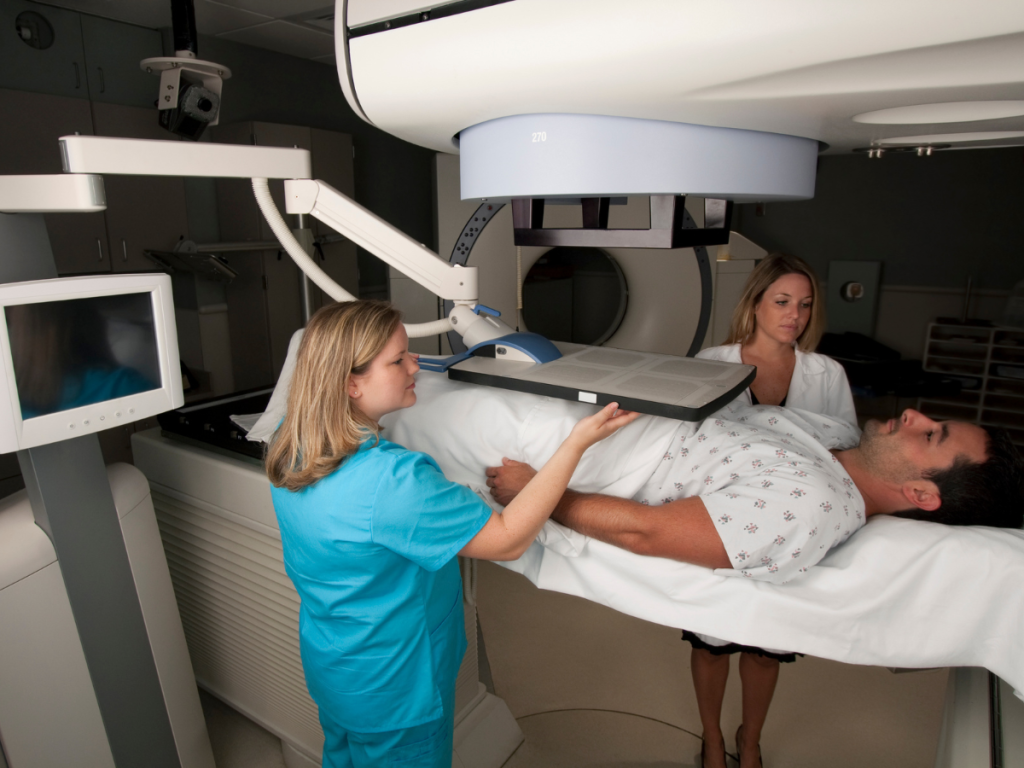
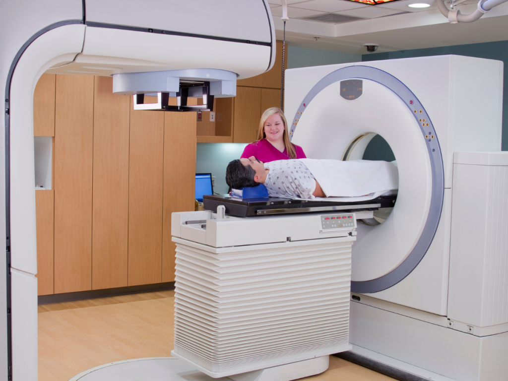
Imaging techniques play a crucial role in diagnosing and staging prostate cancer. Different types of scans are utilized to visualize the prostate gland and surrounding tissues, helping to assess the extent and spread of the cancer. Prostate scans commonly used include:
Transrectal Ultrasound (TRUS)
Transrectal ultrasound involves the use of a small probe inserted into the rectum to produce detailed images of the prostate. It helps in identifying any suspicious areas or abnormalities within the prostate gland.
Magnetic Resonance Imaging (MRI)
MRI uses powerful magnets and radio waves to create detailed images of the prostate and surrounding tissues. It provides clearer visualization of the prostate and helps assess the extent of cancer involvement.
Computed Tomography (CT) Scan
CT scans use X-rays and computer technology to create cross-sectional images of the body. They are often used to determine if cancer has spread to nearby lymph nodes or other organs.
Bone Scan
A bone scan is conducted to evaluate if prostate cancer has spread to the bones. It involves injecting a small amount of radioactive material into the bloodstream, which is detected by a special camera to identify areas of abnormal bone activity.
Staging of Prostate Cancer
Staging refers to determining the extent and spread of cancer within the body. The most commonly used staging system for prostate cancer is the TNM system, which considers the size of the tumor (T), lymph node involvement (N), and the presence of distant metastasis (M). Imaging tests, including prostate scans, play a crucial role in accurately staging prostate cancer.

It’s important to note that the specific tests and imaging techniques used may vary depending on individual cases and healthcare provider preferences. The healthcare team will consider various factors, including the patient’s age, overall health, and specific characteristics of the cancer, to determine the most appropriate diagnostic and staging approach.
If you have concerns about prostate cancer or have been advised to undergo diagnostic tests, it is essential to consult with a qualified healthcare professional. They will evaluate your individual situation, explain the different tests available, and guide you through the diagnostic process.
In conclusion, diagnosing and staging prostate cancer involve a combination of tests and imaging techniques.
