Understanding the Benefits of Hernia Ultrasound: A Comprehensive Guide
Hernias are a common medical condition affecting millions of individuals worldwide. Traditional methods of diagnosing hernias include physical examinations and imaging techniques like X-rays and CT scans. However, in recent years, ultrasound has emerged as a valuable tool in the diagnosis and monitoring of hernias. In this comprehensive guide, we will explore the benefits of hernia ultrasound and how it revolutionizes the way healthcare professionals diagnose and treat hernias.
- The Basics of Hernia Ultrasound:
Hernia ultrasound, also known as sonography, utilizes sound waves to create real-time images of the inside of the body. It is a safe and non-invasive imaging technique that can effectively diagnose different types of hernias, including inguinal, umbilical, and incisional hernias. By capturing detailed images of the affected area, ultrasound helps physicians make accurate diagnoses, assess the severity of the condition, and plan effective treatment strategies.
- Advantages of Hernia Ultrasound over Traditional Imaging Techniques:
2.1 Enhanced Diagnosis Accuracy:
One of the key benefits of hernia ultrasound is its ability to provide detailed and high-resolution images of the hernia. Unlike X-rays and CT scans, ultrasound does not use ionizing radiation, making it safer for patients, especially pregnant women and children. The real-time images obtained through ultrasound help physicians identify the exact location, size, and type of hernia, leading to accurate diagnoses and appropriate treatment plans.
2.2 Cost-Effectiveness:
Compared to other imaging techniques, hernia ultrasound is relatively more cost-effective. It eliminates the need for expensive equipment and radiation exposure, resulting in lower healthcare costs for both patients and healthcare providers. Additionally, ultrasound examinations can be performed in outpatient settings, reducing the need for hospital admissions and further lowering healthcare expenses.
2.3 Real-Time Monitoring:
Hernia ultrasound offers the advantage of real-time monitoring during and after surgical procedures. Surgeons can visualize the hernia repair process, ensuring accurate placement and secure fixation of the mesh or sutures. This real-time monitoring facilitates immediate adjustments if needed, leading to improved surgical outcomes and reduced postoperative complications.
- Hernia Ultrasound Procedure:
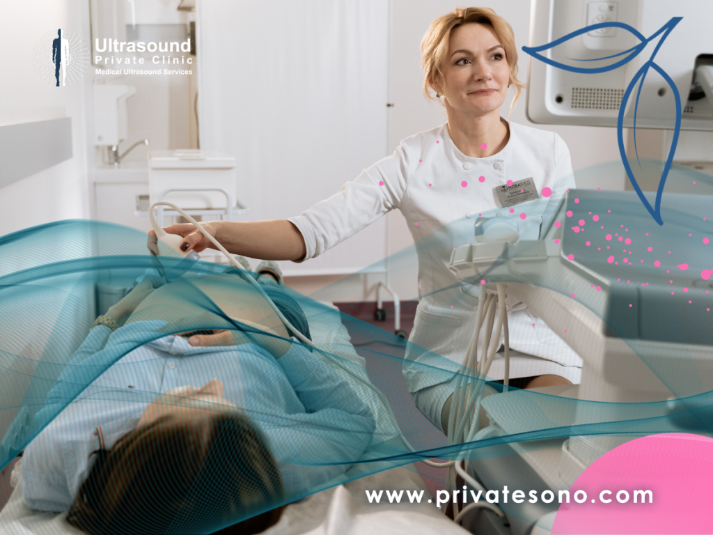
The hernia ultrasound procedure is simple, painless, and quick, typically taking less than 30 minutes. Here is a step-by-step overview of what patients can expect during a hernia ultrasound examination:
Step 1: Preparation – Patients may be asked to wear loose-fitting clothing and remove any metallic objects or jewelry around the area being scanned.
Step 2: Positioning – The patient will be asked to lie down on an examination table, exposing the area of concern.
Step 3: Gel Application – A clear gel is applied to the skin to improve sound wave transmission between the transducer and the body.
Step 4: Transducer Movement – The ultrasound technician moves the transducer (a handheld device) over the skin in the area of the suspected hernia. The transducer emits sound waves and collects the echoes, which are then converted into real-time images on a monitor.
Step 5: Image Analysis – The physician carefully analyzes the obtained images, looking for any signs of hernia, such as a bulge or weakened abdominal wall.
- The Role of Hernia Ultrasound in Treatment Planning:
Hernia ultrasound not only diagnoses hernias but also aids in treatment planning. The obtained images help surgeons determine the most appropriate surgical approach, such as open repair or laparoscopic surgery. Additionally, ultrasound is a valuable tool for assessing the viability of herniated organs or tissues, guiding surgeons in making informed decisions during surgery. This minimizes the risk of complications and improves patient outcomes.
- Conclusion and Call to Action:
In conclusion, hernia ultrasound has revolutionized the way hernias are diagnosed and treated. Its accuracy, cost-effectiveness, and real-time monitoring capabilities make it a superior imaging technique compared to traditional methods. If you suspect you have a hernia or have been diagnosed with one, consider discussing the benefits of hernia ultrasound with your healthcare provider.


You really make it appear really easy with your presentation however I to find this topic to be really something which I feel I might by no means understand. It seems too complex and very wide for me. I’m taking a look ahead in your next publish, I’ll attempt to get the hang of it!