Abdomen & Pelvic Ultrasound Scan

Ultrasound Abdomen & Pelvic - Price: £399
An Abdomen and Pelvic Ultrasound Scan is a safe, painless imaging test that uses sound waves to create detailed pictures of the organs in your abdomen and pelvis. It helps doctors diagnose and monitor various conditions affecting organs like the liver, kidneys, bladder, uterus, ovaries, prostate, and more.
What Does It Evaluate?
- Liver, gallbladder, pancreas, and spleen for signs of disease or damage
- Kidneys and bladder for stones, infections, or abnormalities
- Uterus and ovaries in women to check for cysts, fibroids, or other conditions
- Prostate gland in men for enlargement or abnormalities
- Presence of fluid, masses, or inflammation in the abdominal or pelvic area
Why Might You Need This Scan?
Your doctor may recommend this scan if you experience:
- Abdominal or pelvic pain or swelling
- Unexplained weight loss or bloating
- Changes in urination or bowel habits
- Abnormal blood test results related to liver, kidney, or reproductive organs
- Follow-up for known conditions like gallstones, kidney stones, cysts, or fibroids
- Monitoring during pregnancy or reproductive health assessments
Common Symptoms or Causes
You may need this scan if you notice:
- Persistent abdominal pain or discomfort
- Swelling or a feeling of fullness in your abdomen or pelvis
- Unexplained nausea, vomiting, or jaundice
- Blood in urine or changes in urination
- Irregular menstrual cycles or pelvic pain (in women)
- Difficulty urinating or pelvic discomfort (in men)
Benefits of an Abdomen & Pelvic Ultrasound Scan
- Non-invasive, painless, and radiation-free procedure
- Provides detailed images for accurate diagnosis
- Helps detect and monitor a wide range of conditions early
- Guides treatment plans and follow-up care
- Safe for repeated use and during pregnancy
- Quick and convenient, usually completed within 30 minutes
Concerned about abdominal or pelvic symptoms? Book your Abdomen & Pelvic Ultrasound Scan today.
Book Online Abdomen and Pelvic Ultrasound Scan
If you are unable to make a payment online, please call our office to book your appointment. We’re here to assist you! Tel: 020 7101 3377
Please select a location and time slot to proceed with the booking
If you are unable to make a payment online, please call our office to book your appointment. We’re here to assist you!
Tel: 020 7101 3377
Our Latest Google Reviews
EXCELLENTTrustindex verifies that the original source of the review is Google. Very pleased with my appointment for a knee scan which was done very precisely and slowly and gave a true picture of the whole knee area and back of knee. Was sent the results which was good for me to see for future medication regarding growing arthritis. All staff very helpful with any information you needed. This was the St.Albans branch. Very GoodPosted onTrustindex verifies that the original source of the review is Google. Last minute appointment easily booked the day before. Such a charming doctor and assistant. Very thorough and reassuring and thankfully negative results.Posted onTrustindex verifies that the original source of the review is Google. I needed an immediate scan to put my mind at rest on a significant medical matter. London Private Ultrasound operating from St Albans provided an exemplary customer service. Dr Vakilian was friendly, professional and knowledgeable, ably supported by a charming assistant and receptionist. As well as discussing the scan findings with me during my visit I received a detailed report on the scan with conclusions and recommendations within 24 hours. I strongly recommend London Private Ultrasound to everyone who requires a fast, efficient and courteous medical service.Posted onTrustindex verifies that the original source of the review is Google. Affordable and great service.Posted onTrustindex verifies that the original source of the review is Google. I have previously attended private ultrasound clinics—one was very good, but I also had a negative experience at another clinic in the city, despite paying more. Because of that, I decided to try this location on Welbeck Street after seeing it online, and I had a good feeling from the start. I was fortunate to have Mr. Reza Farahmandfar as my specialist🙏. He performed my breast scan and ultrasound and made me feel completely at ease throughout the appointment. Nothing was too much trouble, and the scan was not painful at all. Everything was calm, smooth, and professional, and his gentle approach was truly reassuring. Thank you for taking such good care of me and for also delivering the wonderful news that everything is healthy. God bless you and your lovely family.🙏🙏🙏🌷🌷🌷🌺🌺🌺Posted onTrustindex verifies that the original source of the review is Google. Excellent service - friendly and professional, - kept informed throughout and was able to discuss and get helpful answers during procedure. Full results and imaging report sent by next morning following a 5pm scan!! Cannot recommend highly enough. Thank you so much.Posted onTrustindex verifies that the original source of the review is Google. Very professional service with friendly staff would definately recommend.Posted onTrustindex verifies that the original source of the review is Google. Very happy with the service I received here. Mr Farahmandfar was very thorough as well as being friendly and professional. I am really pleased I came and will be back to have regular check ups.Posted onTrustindex verifies that the original source of the review is Google. Excellent service, professional and very quick to get an appointment. Highly recommendPosted onTrustindex verifies that the original source of the review is Google. Easy booking process, with plenty of available appointments. I was seen on time by a very kind and professional doctor who gave me my results immediatly because he knew I was concerned. PDF of the scan images was in my inbox before I'd even left the building!
Private Abdominal & Pelvic Ultrasound in London & St Albans
Same-day abdomen and pelvis ultrasound appointments in London (Harley Street/W1G), St Albans, and Wokingham. If you searched for “private abdominal ultrasound scan,” “ultrasound of abdomen and pelvis,” “pelvic ultrasound near me,” or “scan for pelvis and abdomen,” this page explains what the scans show, how to prepare (fasting/full bladder), how long they take, prices, and how to book.
What does an abdominal ultrasound show?
An abdominal ultrasound surveys the upper and/or lower abdomen. Depending on the referral, we assess:
- Liver & bile ducts: size/texture, focal lesions, biliary dilation; gallbladder for stones, polyps, wall changes.
- Pancreas & spleen: overall morphology (noting that bowel gas can occasionally limit pancreatic views).
- Kidneys & ureters (proximal): stones, obstruction (hydronephrosis), cysts, scarring; bladder as part of lower abdominal review.
- Abdominal aorta & major vessels: screening for aneurysm (AAA) and measuring vessel calibre; optional Doppler.
- Anterior abdominal wall: lumps/bumps, hernias when requested.
Lower abdominal views often include bladder; in women, pelvic organs are best assessed under the pelvic section below.
What does a pelvic ultrasound show?
A pelvic ultrasound evaluates the pelvic organs. We tailor to sex and symptoms:
- Female pelvis (transabdominal ± transvaginal): uterus (size/shape), endometrium (thickness/contour), ovaries (follicles/cysts), adnexa, free fluid. Optional 3D/TVS for fibroids, polyps, adenomyosis, IUD position, and fertility mapping.
- Male pelvis (transabdominal): bladder, prostate region (size/indentation on bladder), seminal vesicles when visible; scrotal/testicular ultrasound can be added if indicated.
Pelvic ultrasound helps investigate pelvic pain, abnormal bleeding, suspected ovarian cysts, fibroids, PCOS morphology, early pregnancy queries, urinary symptoms, and to follow up known conditions.
Combined abdomen & pelvis scan
A combined abdomino-pelvic ultrasound reviews the upper abdominal organs and the pelvic organs in one visit—useful for non-specific pain, bloating, weight loss, abnormal blood tests, or when both hepatobiliary and gynaecology/urinary systems need assessment. We can include mesenteric fat thickness measurement, renal/bladder review, and targeted lump/hernia checks on request.
Preparation: fasting & full bladder
- Abdomen: Fast from food for 6 hours (water allowed) to reduce bowel gas and optimise the gallbladder. Avoid fizzy drinks and heavy coffee beforehand.
- Pelvis (female & male): Arrive with a comfortably full bladder (drink ~500–750 ml water 45–60 minutes before; don’t empty). A full bladder acts as an acoustic window.
- Transvaginal (TVS): You’ll be asked to empty your bladder just before the internal scan.
- Periods: Non-urgent endometrial assessment is best after bleeding; urgent pain/bleeding assessments can be performed during menses.
- Medication: Continue as prescribed unless advised otherwise.
How the scan is performed
- Brief history: Symptoms, duration, cycle day (if applicable), prior imaging.
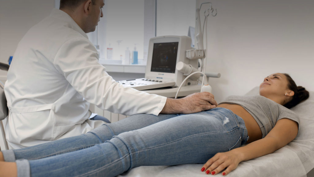
- Transabdominal scan: Gel on the tummy; we scan the organs in real time and take measurements.
- Pelvic component: Transabdominal for all; TVS offered to women for higher resolution (consent, chaperone available).
- Doppler/3D when indicated: Vascular flow or uterine cavity detail can be added where clinically helpful.
- Feedback: Immediate verbal summary; structured written report with images follows.
Common reasons to have an abdominal or pelvic ultrasound
Abdomen
- Upper/right-sided abdominal pain; suspected gallstones or biliary colic
- Abnormal LFTs, fatty liver follow-up, AAA screening
- Kidney pain, stones, UTIs, hydronephrosis
- Palpable lump/hernia, abdominal wall pain
- Non-specific pain, bloating, weight change
Pelvis (Female)
- Pelvic pain, painful periods, heavy or irregular bleeding
- Suspected ovarian cysts, fibroids, polyps, adenomyosis
- Fertility & pre-conception checks; follicle tracking
- Early pregnancy reassurance when clinically indicated
- IUD position, post-operative follow-up
Pelvis (Male)
- Urinary frequency, bladder emptying issues
- Prostate impression on bladder (TA proxy)
- Pelvic/groin pain; add scrotal US if needed
Results & example report wording
- Normal abdomen: “Liver normal in size/echotexture; gallbladder thin-walled, no stones; bile ducts not dilated; pancreas and spleen within normal limits; kidneys normal, no hydronephrosis; aorta normal calibre.”
- Gallstones: “Multiple echogenic foci with posterior acoustic shadowing in gallbladder; no wall thickening or pericholecystic fluid; CBD x mm (within normal range).”
- Normal female pelvis: “Uterus normal in size; endometrium smooth (x mm, phase-appropriate); ovaries with follicles; no adnexal mass or free fluid.”
- Ovarian cyst: “Right ovarian anechoic cyst 3.2 cm, thin wall, no internal solid components or Doppler flow—features of a simple cyst.”
- Fibroids: “Intramural fibroids in anterior wall up to 2.8 cm; cavity not significantly distorted on TA views.”
Prices
- Abdominal ultrasound: typically £140–£220 (scope-dependent)
- Pelvic ultrasound (TA ± TVS): typically £140–£240 (female/male protocols)
- Combined abdomen & pelvis: typically £220–£320
- Add-ons on request: mesenteric fat thickness, hernia review, vessel Doppler, 3D/TVS gynae protocol
Fees include the scan, same-day verbal summary, and a consultant-approved written report with key images.
Locations & “near me”
We offer same-day/next-day appointments in:
- Central London (Harley Street/W1G incl. Welbeck St.) – abdomen, pelvis, combined, Doppler, and mesenteric fat thickness.
- St Albans – adult & child scans; evening/weekend slots.
Frequently asked questions
Is a pelvic ultrasound the same as an abdominal ultrasound?
No. Abdominal ultrasound assesses upper abdominal organs (liver, gallbladder, pancreas, spleen, kidneys, aorta). Pelvic ultrasound focuses on uterus/endometrium/ovaries in females and bladder/prostate region in males. They are often combined when symptoms overlap.
Do I need to fast or have a full bladder?
For the abdomen, fast for ~6 hours (water allowed). For the pelvis, arrive with a comfortably full bladder. If we then perform an internal (TVS) scan, you will empty your bladder first.
Can I have a pelvic scan during my period?
Yes for urgent problems. For non-urgent endometrial assessment, post-menses is usually clearer. TVS is optional and performed with consent.
How long do the scans take and when do I get results?
Abdominal or pelvic scans usually take 15–25 minutes; combined 25–40 minutes. You’ll receive a same-day verbal summary and a written report with images shortly after.
What can abdominal/pelvic ultrasound detect?
Common findings include gallstones, bile duct dilation, fatty liver features, kidney stones or obstruction, abdominal wall hernias/lumps, ovarian cysts, fibroids, endometrial polyps, simple pelvic fluid, and bladder abnormalities. Some conditions (e.g., subtle bowel disease, small deep endometriosis) may require other tests such as MRI/CT or endoscopy.
Is ultrasound good for cancer detection?
Ultrasound can detect many masses, but it is not a comprehensive cancer screen. If we see a suspicious feature, we will recommend the appropriate next step (e.g., further imaging or referral).
What about mesenteric fat thickness?
On request, we can measure mesenteric/visceral fat thickness as part of metabolic risk discussions. It’s an optional add-on and interpreted alongside clinical context.
Book your scan
Choose abdominal, pelvic, or combined scans. Same-day and next-day appointments are often available.
Private Ultrasound Clinic
All part of our services, from our specialists to our technology and, of course, our clinic, is designed to deliver the greatest possible experience for all of our patients and visitors.
We are conveniently located a stone throw famous Harley Street of London and our clinic is a place where you may feel safe and clean, comfortable, and reassuring environment.
Central London Branch: 27 Welbeck Street, London, W1G 8EN
St Albans Branch : 54-56 Victoria St, St Albans, AL1 3HZ
Tel: 020 7101 3377


