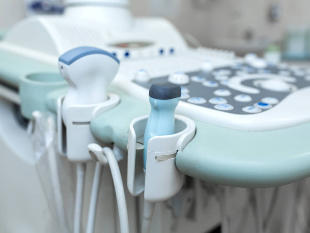
Prostate health is a crucial aspect of men’s overall well-being, and regular screenings play a vital role in detecting potential issues early on. One commonly used diagnostic tool is a prostate ultrasound. In this blog post, we will explore what a prostate ultrasound entails, its significance in prostate health, and how the procedure is performed. So, let’s dive in!
Understanding Prostate Ultrasound
A prostate ultrasound is a non-invasive imaging technique that utilizes high-frequency sound waves to create detailed images of the prostate gland. It provides valuable insights into the size, shape, and condition of the prostate, aiding in the diagnosis of various prostate-related conditions.
The Importance of Prostate Ultrasound
Prostate ultrasound is commonly used to evaluate and monitor conditions such as prostate cancer, benign prostatic hyperplasia (BPH), prostatitis, and other abnormalities. By visualizing the prostate gland, doctors can assess its structure, identify any abnormalities, and determine the appropriate course of action for further treatment or monitoring.

How Prostate Ultrasound is Performed:
Preparation: Prior to the procedure, patients may be instructed to drink fluids and refrain from emptying their bladder to ensure better visibility during the ultrasound. It is advisable to follow the specific guidelines provided by the healthcare professional.
Positioning: The patient typically lies on their side or back, with their knees bent and pulled toward the chest. This position allows for easier access to the prostate gland.
Transrectal Ultrasound: A lubricated ultrasound probe, known as a transducer, is gently inserted into the rectum. The transducer emits sound waves that bounce off the prostate gland, creating real-time images on a monitor.
Image Capture: The ultrasound technician or radiologist moves the transducer within the rectum to capture images of different sections of the prostate gland. This process may take a few minutes to complete.
Post-Procedure: Once the images are obtained, the transducer is removed, and the procedure is complete. Patients can resume their normal activities immediately after the ultrasound.
Benefits and Considerations
Prostate ultrasound offers several advantages, including its non-invasive nature, real-time imaging capabilities, and the ability to visualize the prostate gland directly. However, it is important to note that prostate ultrasound alone may not provide a definitive diagnosis. In some cases, additional tests such as prostate biopsy or blood tests may be necessary to confirm or rule out certain conditions.

Conclusion
Prostate ultrasound is a valuable diagnostic tool in assessing prostate health and detecting potential abnormalities. By providing detailed images of the prostate gland, this procedure aids in the early detection and management of various prostate-related conditions. If you have concerns about your prostate health, consult with your healthcare provider, who can guide you on the appropriate screenings and examinations, including a prostate ultrasound if needed.
Remember, proactive monitoring and early detection are crucial in maintaining optimal prostate health, so prioritize regular check-ups and discussions with your healthcare professional.
